
Skeletal muscle is built from a nested hierarchy of structures, each wrapped in its own connective tissue, and it contracts through a precise chain of electrical and chemical events called excitation-contraction coupling.
Structure Hierarchy
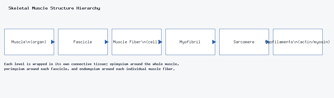
From largest to smallest: muscle (the whole organ) → fascicle → muscle fiber (the individual cell) → myofibril → sarcomere → myofilaments (actin and myosin).
Connective Tissue Wrappings
- Epimysium — surrounds the entire muscle.
- Perimysium — surrounds each fascicle.
- Endomysium — surrounds each individual muscle fiber.
Filament Types
Thin filaments are made of three proteins working together: actin, the structural backbone that myosin binds to; tropomyosin, which physically blocks myosin’s binding sites on actin at rest; and troponin, which shifts tropomyosin out of the way when it’s activated by a rush of calcium.
Thick filaments are made of myosin, a motor protein. Each myosin head has both an actin-binding site and an ATP-binding site — when both are filled, the myosin head stretches into position to bind F-actin.
The Troponin-Tropomyosin Complex
At rest, tropomyosin blocks myosin’s binding sites on actin, so no contraction can happen. Upon stimulation, Ca²⁺ binds troponin, which pulls tropomyosin out of the way, letting myosin bind actin and drive contraction.
Calcium Control
Calcium is stored in the sarcoplasmic reticulum (SR). It’s released through voltage-gated Ca²⁺ channels the moment an action potential arrives, and afterward it’s actively pumped back into the SR to let the muscle relax.
Excitation-Contraction Coupling
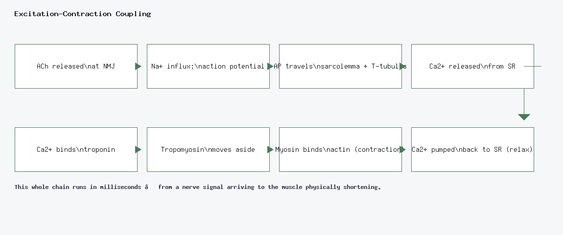
- A motor neuron releases acetylcholine (ACh) at the neuromuscular junction (NMJ).
- ACh binds its receptors, and Na⁺ floods the muscle fiber, creating an action potential.
- The action potential travels along the sarcolemma and down the T-tubules.
- Voltage-gated Ca²⁺ channels open in the sarcoplasmic reticulum, and Ca²⁺ floods the cytosol.
- Ca²⁺ and ATP binding to troponin drives contraction.
- Ca²⁺ is pumped back into the SR, and the muscle relaxes.
Force Generation
Most everyday force is generated through isometric contraction — tension without a change in muscle length. As shortening velocity increases, the force a muscle can produce actually decreases; the two are inversely related. Tension is strongest when there’s maximum overlap between actin and myosin filaments — too little overlap or too much overlap (filaments jammed together) both reduce the force a sarcomere can generate.
Levers in the Body
- 1st class — the neck (head tilting or nodding).
- 2nd class — a calf raise (the triceps surae acts here).
- 3rd class — a biceps curl.
Tetanus (Continuous Contraction)
Muscular tetanus — a sustained, unrelaxing contraction — is caused by high-frequency stimulation, or by a toxin or drug that prevents Ca²⁺ reuptake or keeps Na⁺ channels from inactivating.