
The autonomic nervous system (ANS) runs the body’s background operations — heart rate, digestion, airway diameter — without any conscious input. It works through two divisions that usually pull in opposite directions on the same organs.
The Two-Neuron Chain
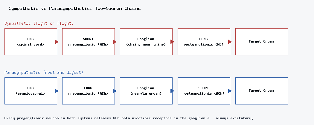
Both the sympathetic and parasympathetic divisions work through the same basic structure: a preganglionic neuron runs from the CNS to a ganglion, then a postganglionic neuron runs from that ganglion to the target organ. The synapse between the two always happens inside the ganglion. The hypothalamus is the master control center for the whole ANS, while the medulla oblongata handles moment-to-moment autonomic life functions like breathing and heart rate.
Sympathetic (Fight or Flight)
The alert response, originating from the spinal cord. Preganglionic fibers are short, postganglionic fibers are long.
- Sympathetic chain ganglia — run parallel to the spine.
- Collateral ganglia — located in the abdomen.
- Adrenal medulla — releases epinephrine directly into the blood.
Parasympathetic (Rest and Digest)
The relaxation response, originating from the craniosacral region. Here the pattern flips: preganglionic fibers are long, postganglionic fibers are short. Its ganglia sit near or inside the target organs themselves, which is exactly why parasympathetic responses tend to be local and specific rather than body-wide.
Neurotransmitters and Receptors
Preganglionic Neurons (Both Systems)
Every preganglionic autonomic neuron, sympathetic or parasympathetic, originates in the CNS and releases ACh onto nicotinic receptors in the ganglion. Nicotinic receptors are ion channels that always depolarize — they’re always excitatory, with no exceptions.
Postganglionic Neurons (Where They Differ)
The postganglionic neuron receives that ACh signal and sends its own axon out to the target organ — but what it releases, and what kind of receptor it acts on, depends on which division it belongs to.
Muscarinic receptors (parasympathetic) — the postganglionic neuron releases ACh onto these G-protein coupled receptors, which can be excitatory or inhibitory but act slowly. Examples: slowing heart rate, or constricting airways.
Adrenergic receptors (sympathetic) — the postganglionic neuron releases norepinephrine (NE) or epinephrine onto these receptors:
- Alpha 1 → vasoconstriction.
- Beta 1 → increases heart rate and contractility.
- Beta 2 → bronchodilation, vasodilation.
A quick reminder of why this all matters clinically: epinephrine and albuterol are chemically related but act on different receptor sites — which is exactly why an asthma inhaler (a beta-2 selective drug) doesn’t send your heart racing the way a shot of epinephrine would.
Sympathetic vs. Parasympathetic Involvement
All ANS ganglia receive ACh from their preganglionic neurons, but the sympathetic response lasts longer overall, since norepinephrine has a longer half-life than ACh. Most organs receive dual innervation — input from both divisions pulling in opposite directions:
- Heart rate — sympathetic increases it, parasympathetic decreases it.
- GI tract — sympathetic slows digestion, parasympathetic speeds it up.
Short vs. Long Reflexes
- Short reflexes — bypass the CNS entirely, making them faster.
- Long reflexes — route through the CNS, which is slower but allows for more coordination.
Both are controlled mostly by the spinal cord, hypothalamus, and brainstem — none of it is consciously directed.