
The central nervous system (CNS) — brain and spinal cord — is protected by bone, cushioned by cerebrospinal fluid, and organized into distinct regions that each handle a different piece of the job: movement, sensation, homeostasis, and everything in between.
Glial Cells
Glial cells support neurons rather than transmitting signals themselves — think of them as the neuron’s maintenance crew.
- Astrocytes — tightly wrap around brain capillaries to help form the blood-brain barrier, letting sodium and potassium through while blocking harmful substances.
- Oligodendrocytes (CNS) — large cells near the neuron whose long processes each cover multiple segments of axon, forming the myelin sheath that speeds up signal propagation.
- Schwann cells (PNS) — do the same myelinating job as oligodendrocytes, but each Schwann cell wraps only one segment of one axon.
- Ependymal cells — produce, secrete, and circulate cerebrospinal fluid (CSF).
- Microglia — the brain’s resident immune cells; clear pathogens and dead cells.
Main Brain Structures
Cerebrum
The largest portion of the brain, split into 2 hemispheres (left and right) and 4 lobes (frontal, parietal, temporal, occipital). Two strips across the cerebrum matter most for exams: the precentral gyrus (primary motor cortex) sends signals to voluntary muscles, and the postcentral gyrus (primary sensory cortex) receives signals from the body’s sensory receptors.
Cerebellum
Sits below the occipital lobe toward the back of the brain. Handles balance, posture, and coordination of voluntary movement, using input from the motor cortex and from proprioceptors around the body.
Diencephalon
- Thalamus — the relay center for sensory and motor information; it sorts and routes signals to where they need to go, almost like a switchboard.
- Hypothalamus — maintains homeostasis: controls the ANS, hormone regulation, temperature, hunger, and circadian rhythm.
- Pineal gland (epithalamus) — regulates sleep and wakefulness by producing melatonin.
Brain Stem
- Pons — the communication bridge between the cerebrum and cerebellum.
- Medulla oblongata — executes autonomic life functions, relayed via the thalamus.
- Midbrain — controls auditory and visual reflexes, and produces dopamine, which governs risk/reward processing.
Other Notable Areas
- Broca’s area — in the left frontal lobe; governs speech production (the motor side of speech).
- Wernicke’s area — in the posterior left hemisphere; governs speech comprehension (understanding language).
- Ventricles — cavities inside the brain that circulate cerebrospinal fluid.
Limbic System
Links conscious and unconscious processing — emotion and the body’s responses to it — largely through the hypothalamus. The amygdala responds to fear; the hippocampus is central to forming memories.
Meninges and Cerebrospinal Fluid
The meninges are the protective connective tissue layers wrapping the brain and spinal cord, from outside in: skull bone, dura mater (protective outer layer), the dural venous sinus (where venous blood flows and CSF drains in from the arachnoid villi), the subarachnoid space (circulates CSF, and needs to stay at high pressure to keep pushing CSF into the blood), and finally the pia mater, the innermost layer directly covering the brain.
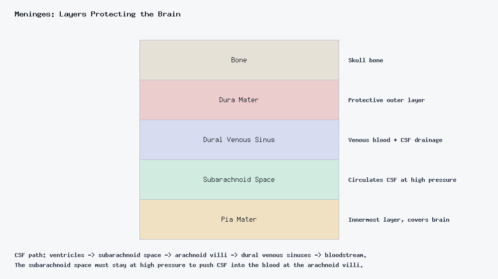
Cerebrospinal fluid (CSF) is a colorless fluid that circulates through the brain’s ventricles. It provides shock absorption, buoyancy (reducing the effective weight of the brain by about 95%), and it circulates nutrients while clearing waste. Its path: ventricles → subarachnoid space → arachnoid villi → venous sinuses.
Blood-Brain Barrier
Formed by astrocytes together with tight junctions between endothelial cells. It freely passes O₂, CO₂, small lipids, and alcohol, while restricting proteins, toxins, and most drugs — which is exactly why so few medications are able to reach the brain directly.
Spinal Cord
The spinal cord is the main communication line between the brain and the rest of the body.
Gray Matter
Made of neuron cell bodies and short, unmyelinated neurons. In cross-section it’s shaped like an H, with anterior, posterior, and lateral horns.
- Anterior horn — houses the cell bodies of lower motor neurons (LMNs). Their axons exit through the ventral roots to innervate skeletal muscle — this is the motor side.
- Posterior horn — contains interneurons that receive sensory input. Sensory neurons enter through the dorsal roots and synapse here — this handles touch, pain, temperature, pressure, and proprioception.
White Matter
Mostly myelinated axons (with some unmyelinated ones mixed in), organized into ascending (sensory) and descending (motor) tracts.
Major Tracts
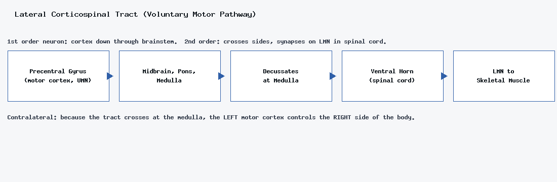
Lateral corticospinal tract (motor) — carries voluntary commands from the brain to skeletal muscle. The 1st order neuron travels from the precentral gyrus (motor cortex, upper motor neuron) down through the midbrain, pons, and medulla oblongata, where the fibers decussate (cross sides). The 2nd order neuron picks up at the ventral horn of the spinal cord and synapses onto the lower motor neuron (LMN), which connects to skeletal muscle fibers. Because the tract crosses in the medulla, the pathway is contralateral — the left motor cortex ultimately controls the right side of the body.
Lateral spinothalamic tract (sensory: pain and temperature) — runs PNS to CNS. The 1st order neuron carries the signal from the sensory receptor through the dorsal root ganglion, synapsing at the posterior dorsal horn. The 2nd order neuron crosses to the opposite side right there and ascends the spinal cord to the medulla. The 3rd order neuron continues to the thalamus, then on to the postcentral gyrus (primary sensory cortex).
Dorsal column-medial lemniscus tract (sensory: fine touch, vibration) — also PNS to CNS, but crosses later. The 1st order neuron ascends ipsilateral (same side) up the spinal cord and synapses in the medulla oblongata. The 2nd order neuron crosses (decussates) there and synapses with the thalamus. The 3rd order neuron continues from the thalamus to the primary sensory cortex.
The key difference to remember: the spinothalamic tract crosses immediately at the spinal cord, while the dorsal column-medial lemniscus tract travels up the same side and only crosses once it reaches the medulla. That’s also why upper motor neurons (UMN) refer only to the CNS portion of a motor pathway (from the precentral gyrus down), while lower motor neurons (LMN) refer to the CNS-to-PNS-to-muscle portion.
Reading Injury Symptoms by Location
- Brain damage — pain, temperature, and touch signals come from contralateral receptors, so damage to the right side of the brain shows up as sensory loss in the left foot, for example.
- Spinal cord damage — pain and temperature travel on the opposite side of the body from the affected location, since the spinothalamic tract has already crossed by that point.
- Spinal nerve damage — all sensory and motor signals on the same side of the body are affected, but only for the specific nerves involved (nothing has crossed yet at this level).