
The peripheral nervous system (PNS) is everything outside the brain and spinal cord — the nerves that connect the CNS to the rest of the body, plus the reflex circuits that let the body react before the brain even gets involved.
PNS Components
Cranial Nerves
Come directly from the brain or brainstem, and can be sensory, motor, or mixed. Several also carry parasympathetic fibers.
Spinal Nerves
All spinal nerves are mixed (both sensory and motor), and they come off the spinal cord in regions:
- Sympathetic spinal nerves — thoracic (T1–T12) innervate the chest wall; lumbar (L1–L5) innervate the lower back and anterior leg.
- Parasympathetic spinal nerves — sacral (S2–S4) innervate pelvic organs, the bladder, and the lower digestive tract.
- Cervical nerves (C1–C8, sensory and motor) — serve the neck, shoulders, and arms.
- A worthwhile distinction: sacral nerves include S2–S4 (parasympathetic), but S1 is not parasympathetic — it’s easy to lump the whole sacral region together, but S1 is the exception.
Nerve Anatomy
The connective tissue wrappings of a peripheral nerve mirror the ones seen in skeletal muscle:
- Epineurium — wraps the whole nerve (“epi-” means closer to the surface).
- Perineurium — wraps each fascicle.
- Endoneurium — wraps the actual individual axon fibers.
Two more directional terms worth locking in: dorsal / afferent refers to sensory signals; ventral / efferent refers to motor signals. Axons are what physically connect a sensory or motor receptor to the dorsal or ventral root ganglia.
Reflexes
Reflexes never require conscious thought — they’re driven by the spinal cord, hypothalamus, and brainstem. At minimum, a reflex needs a sensory neuron and a lower motor neuron; an interneuron is only involved in some reflex types.
Monosynaptic Stretch Reflex
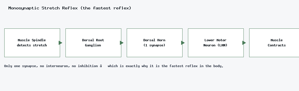
The muscle spindle detects a stretch, and a 1st order sensory neuron carries the signal through the dorsal root ganglion to the dorsal horn, where a single synapse connects directly onto the LMN, causing contraction. Because there’s no interneuron and no inhibition of another pathway, this is the fastest reflex in the body — and it always causes contraction, never relaxation.
Reciprocal Inhibition
Take the quads and hamstrings as an example: the sensory neuron from the quad’s muscle spindle synapses onto the LMN (causing quad contraction) and onto an inhibitory interneuron, which inhibits the hamstring from contracting at the same time — producing an IPSP in the antagonist muscle. This is the fastest reflex for muscle relaxation, but it’s slower than the monosynaptic reflex, since it requires an extra synapse and an interneuron. In short: sensory neuron → inhibitory interneuron → antagonist inhibited.
Crossed Extensor Reflex
Think of accidentally stepping on glass: in the injured leg, the pain signal travels the spinothalamic pathway, flexors contract, and extensors relax (pulling the foot up). In the opposite leg, extensors contract to keep balance on one foot. This is slower than the other reflexes, since it involves multiple interneurons and a contralateral crossing of the signal. It’s polysynaptic, and results in the opposite leg extending for balance.
Golgi Tendon Reflex
A protective, polysynaptic reflex that prevents muscle and tendon damage from excessive force by causing the muscle to relax when tension gets too high. Excessive tension activates Golgi tendon receptors, and the stimulus travels through the dorsal root into the spinal cord; the sensory neuron also synapses onto an inhibitory interneuron there. It’s a 2-synapse reflex, the fastest relaxation reflex the body has for protecting a tendon from excessive tension.
Sensory and Motor Signal Pathways
Getting a sensation to the CNS starts with a graded potential at the receptor, which — if strong enough — becomes an action potential. Getting a motor command back out to the body follows the reverse route. One more concept worth knowing: adaptation, where a receptor’s response weakens over time under constant stimulation (which is why you eventually stop noticing your clothes against your skin).
Neuromuscular Junction Toxins
- Botox — prevents ACh release into the neuromuscular junction. No ACh means no end-plate potential, which means no muscle action potential — leading to flaccid paralysis.
- Succinylcholine — acts like ACh and binds nicotinic receptors, opening Na⁺ channels and causing an initial depolarization (a brief contraction) — but it doesn’t detach, so the receptor stays open. Since the channels never reset, no further action potential can occur, and the result is flaccid paralysis.
Both toxins end in the same outcome — flaccid paralysis — but for opposite reasons: one blocks ACh from ever binding, the other locks the receptor open so it can never reset.